< Perspectives
Stents under strain: Why device choice matters more than ever

A study published last week in the International Journal of Gastrointestinal Intervention has highlighted how oesophageal stents are subject to progressive in vivo corrosion and mechanical failure. This probably won’t come as much of a surprise – stent failures are certainly not unheard of, but the research conducted at the University of Chester shows that the materials used in the construction of stents may matter more than we previously thought. For interventional radiologists, the message is clear; as patient survival improves, stent durability is becoming a critical determinant of long-term outcomes.

In 2004, survival after oesophageal stent placement in the UK was around three months. Advances in oncological and palliative therapies have since extended survival two-to-three-fold, with five-year survival for oesophageal cancer in England approaching 20% in 2023. As a result, stents are remaining in situ for longer, and failure is being seen more often.
Gastric acid exposure is likely the main factor driving stent corrosion; with 60–70% of oesophageal stents being placed over the GOJ, contact with hydrochloric acid is unavoidable. Over time, this appears to degrade the nitinol wires forming the stent skeleton, reducing radial expansion force and making it more susceptible to fracture. The quality of the raw nitinol used has been shown to influence a stent’s chances of fracture, as well as the methods used by the manufacturer to construct the stents and whether the wire skeleton is encapsulated in a polymer.
Gastric acid exposure is likely the main factor driving stent corrosion; with 60–70% of oesophageal stents being placed over the GOJ, contact with hydrochloric acid is unavoidable. Over time, this appears to degrade the nitinol wires forming the stent skeleton, reducing radial expansion force and making it more susceptible to fracture. The quality of the raw nitinol used has been shown to influence a stent’s chances of fracture, as well as the methods used by the manufacturer to construct the stents and whether the wire skeleton is encapsulated in a polymer.

A knitted oesophageal stent retrieved in 3 pieces during a stent revision (Prof. Hans-Ulrich Laasch, The Christie NHS Trust)
The study analysed six stents with varying designs following their removal – scanning electron microscopy (SEM) and tensile testing revealed consistent evidence of corrosion affecting the nitinol mesh. Surface deposits – mainly sodium chloride and carbon-based material – were associated with pitting corrosion and crack formation. Wires showed deformation and brittle fracture (rather than elastic failure). Mechanical testing demonstrated loss of superelasticity and reductions in strength, with some wires even fracturing spontaneously during handling.
Early detection of stent deterioration is essential, as revision is safest and most effective before significant structural collapse occurs. Once integrity is lost, removal may be technically difficult or impossible. Structured follow-up, appropriate acid suppression and clear revision pathways are therefore increasingly important.
Oesophageal stents are no longer short-term palliative devices in most patients. They are longer-term implants in a hostile chemical environment. While radial force and migration rates often remain at the forefront of the conversation, corrosion susceptibility, dwell time expectations and anticipated revisions should all be taken into account. The reality is that all stents are prone to degradation, but some stents degrade a lot faster than others. Choosing your stent carefully and ensuring surveillance is a part of your service will have massive benefits for both you and your patients.
Early detection of stent deterioration is essential, as revision is safest and most effective before significant structural collapse occurs. Once integrity is lost, removal may be technically difficult or impossible. Structured follow-up, appropriate acid suppression and clear revision pathways are therefore increasingly important.
Oesophageal stents are no longer short-term palliative devices in most patients. They are longer-term implants in a hostile chemical environment. While radial force and migration rates often remain at the forefront of the conversation, corrosion susceptibility, dwell time expectations and anticipated revisions should all be taken into account. The reality is that all stents are prone to degradation, but some stents degrade a lot faster than others. Choosing your stent carefully and ensuring surveillance is a part of your service will have massive benefits for both you and your patients.
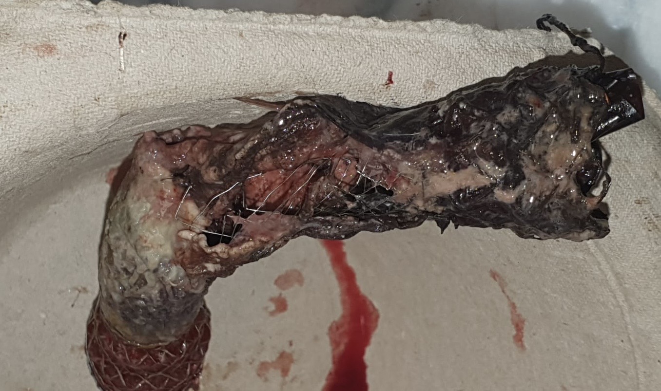
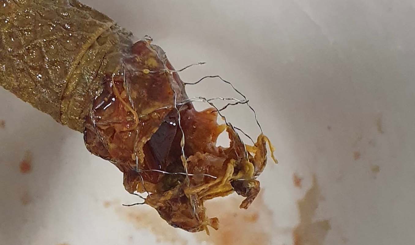
Degraded, fractured stents after removal (Prof. Hans-Ulrich Laasch, The Christie NHS Trust)
Laasch, H.-U., Weaver, C.J., Black, S.J., Smith, G.C. and Edwards, D.W. (2026) ‘In vivo corrosion of esophageal stents: An emerging materials problem’, International Journal of Gastrointestinal Intervention. Published online 24 February 2026


