Attending an assessment clinic for breast.
What to expect

We understand how you might be feeling, and we’re really pleased you’ve found us. We work closely with breast teams across the UK from the Highlands right down to the Channel Islands. They are some of the loveliest people you could be in the care of, and real pros!
Before we discuss what might happen at the assessment clinic, we want to highlight two really important statistics in the hope that it may put your mind at ease a bit:
75% of women recalled for assessment following screening are found to have no breast cancer.1
Breast cancers detected in the early, localised stage have a 5-year survival rate of 99%.2
Preparing for your
assessment appointment
Download checklist >
If the time and date of your appointment is inconvenient please call us and we will do our best to rearrange it for you. Bring somebody with you to keep you company as there can be a lot of waiting. That person can be male or female – they may not be able to come in the room with your for your scans though.
Don’t wear body lotion, spray on deodorant or glitter as this can interfere with the images. Be prepared to have to undress to the waist. It's easier if you wear a skirt or trousers instead of a dress. Wear clothing on your top half that can be easily removed. Finally, be prepared to be here for up to 4 hours for all of your tests – bring a book or a puzzle or something to keep you occupied. For that reason, don’t skip your breakfast! You’re welcome to bring snacks to have in the waiting room – eating and drinking won’t affect any of your tests.
How to prepare

Leave jewellery at home.

What should I wear?
What should I take with me?
- If you take any medications regularly, take these with you or make a note of the names and dosages.
- Something to do – you will probably spend a good amount of time in the waiting room, so take a book, puzzle or something to keep you occupied.
- Some water and a couple of snacks in case you get peckish.
- A pen and paper in case you need to take any notes during your consultation.
- A hair bobble or clip if you have long hair.
- Some handy wipes if you’re self-conscious about not wearing deodorant.
- A supportive friend or family member.
Medical terms you might hear
You might hear some terms you aren’t familiar with while at your assessment appointment. Here are some of the things that might be talked about:
Lesions – A lesion is defined as a region of organ or tissue which has suffered damage or injury through disease. It is a broad term given to any type of abnormal tissue – a cut on your skin or a cyst could be considered a lesion, so don’t worry – it doesn’t necessarily mean the worst. The area of interest in your breast that the clinicians are looking at will likely be referred to as the lesion.
Microcalcifications – Tiny flecks of calcium that can be seen in mammograms. It is not uncommon for breasts to contain a small number of microcalcifications and they are not inherently sinister in themselves (think about it: milk = calcium!) But microcalcifications that appear in some shapes and patterns can be clinically significant. They are very difficult to see under ultrasound because they are so small.
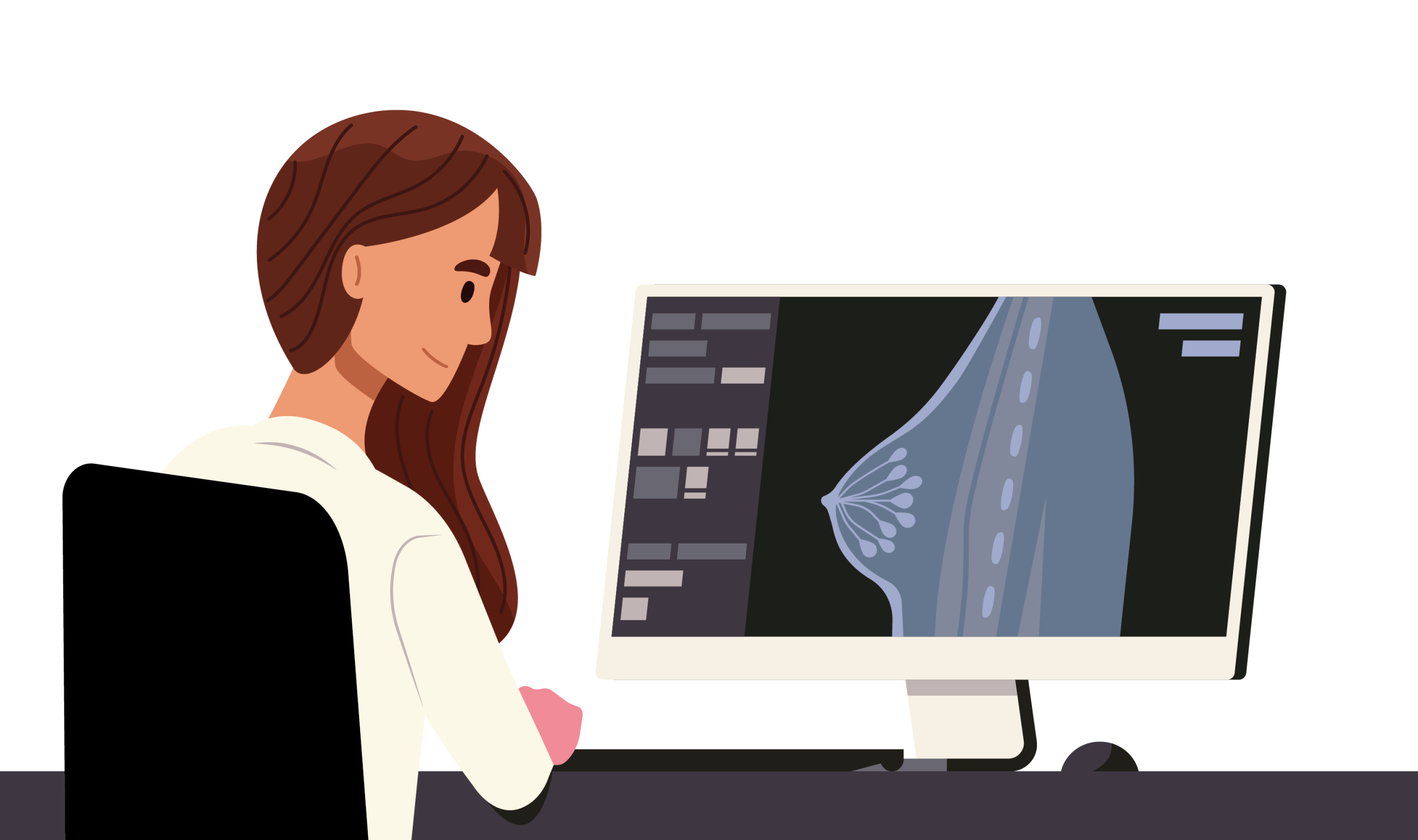
Mammography – X-ray imaging of the breast
Mammogram – The image produced by X-raying the breast
Stereotactic – Using X-ray guidance to perform a procedure
Biopsy – The removal of a small piece of tissue for pathological analysis
Benign – A disease not harmful in effect
Malignant – A disease harmful in effect

Attending Assessment

If you have children, it’s definitely best to leave them at home or arrange for someone to look after them for the duration of your appointment.
If your first language is not English, please note that relatives can’t be used as translators and the hospital needs to arrange for an approved interpreter to attend (this can be done via telephone too). The clinic will need to know about this in advance.
Be aware that there may be some back and forth between the waiting room and the imaging or examination rooms – you may be offered the choice of staying in a hospital gown or redressing to go back into the waiting room.
Additionally, if there is any additional support that you need for your appointment (for instance if you have difficulties with mobility or learning difficulties) do let your breast clinic know beforehand so they can make any necessary accommodations for you.

Assessment Structure
Yes, we need to know about any breast surgery (cosmetic or not), any medications (especially blood thinners) and allergies, if you are breastfeeding or pregnant or if you have a pacemaker or other type of cardiac monitoring device. Knowing your family history is also really helpful. If you are taking any medications, it’s always a great idea to bring it with you in case the breast team need to know your exact medication and dosage.
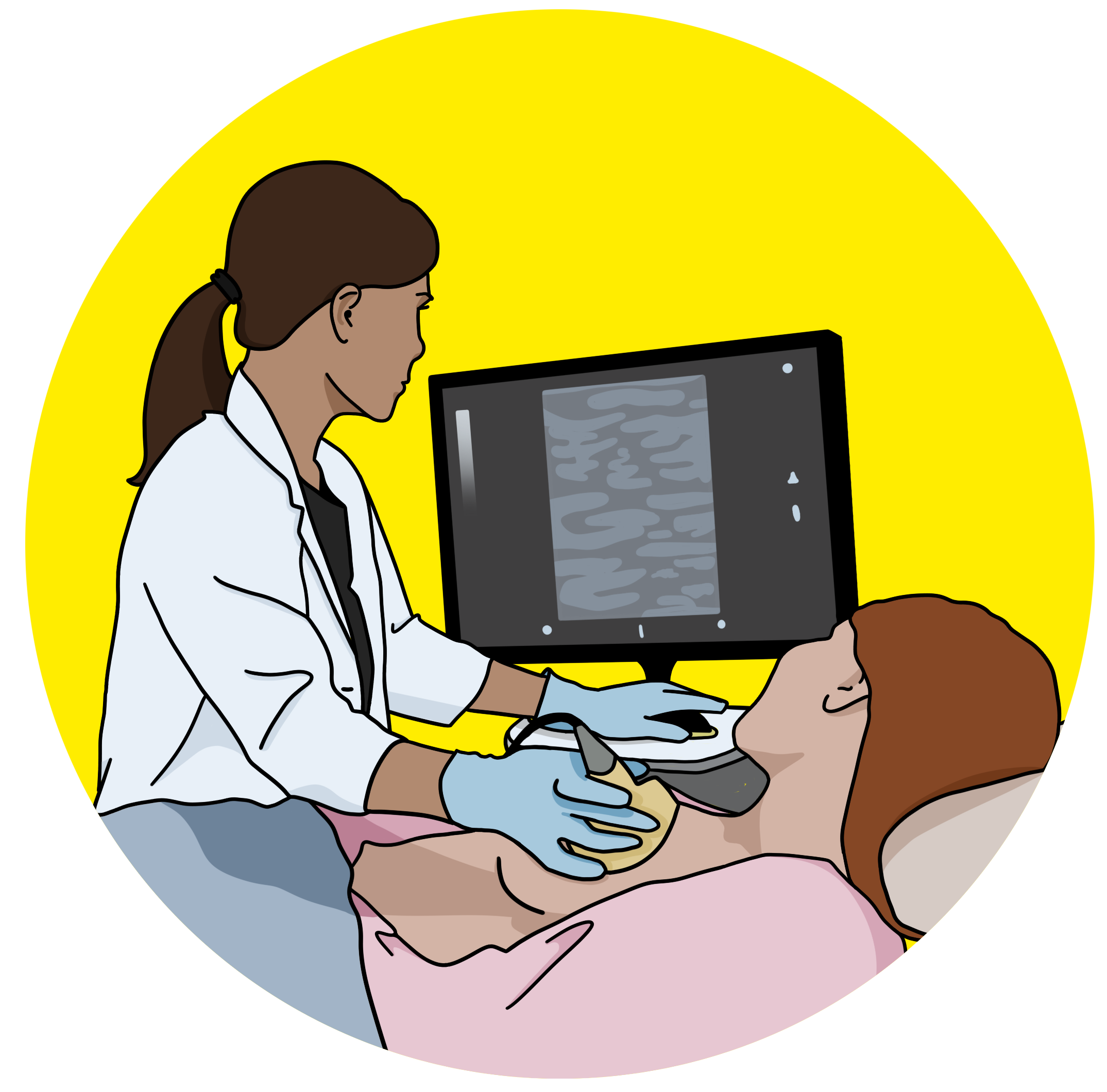
Most people will also undergo an ultrasound scan of the breast and armpit region. If you need a biopsy, the Doctor or Radiographer performing the scan will tell you at this point. If the additional imaging is normal, you will likely be told this and no further testing (including biopsy) will be needed.
You will be invited to an appointment to discuss your results – this should be within 2 weeks of attending clinic. In a small number of cases, a definitive diagnosis cannot be made. Instead these patients are recalled early for further assessment.
“The treatment I received from the breast team was great. Obviously, being extremely anxious having found a lump and not knowing what the next steps were going to be, the team were very caring and considerate.
I was seen first by a consultant who examined me, then I had to wait to have a mammogram. It wasn’t uncomfortable for me and again the radiographer was lovely and reassuring.
After this I was then given an ultrasound. The doctor confirmed it was a cyst and drained it there and then.
The worst part really was the waiting - but I can’t say enough good things about the breast care team!”
It’s completely natural to be nervous about something like this. Do bring someone with you if you can so that you can talk through your anxieties and have someone to take your mind off things while you wait. You can tell your breast care nurse, radiographer or radiologist if you are feeling nervous – let them know what it is you are worried about as they may be able to ease some of your worries. If you do have a lot of questions about your assessment appointment, you are always welcome to call your breast unit before you attend – there will usually be a breast care nurse or radiographer who can talk to you about your concerns – the number to call will be on your appointment letter.
Breast Biopsies
We understand that biopsy is often the most daunting aspect of attending the assessment clinic. We want to break down the biopsy process for you in the hopes that demystifying the process gives you more confidence to attend your appointment.
Core biopsies are performed with special mechanical needles to take a portion of the lesion for testing. The clinician will usually take a few small samples with one of these devices.
VAB biopsies are performed with VAB machines and they use vacuum to pull tissue into the needle. This method could be chosen for several reasons – if you have been recalled due to microcalcifications, as they can be quite scattered, the vacuum helps the clinicians obtain all of the tissue they need for pathology.
Some women have what we refer to as ‘dense breasts’ – this means that they have a lot of glandular tissue, which shows up bright under X-ray (mammography). Younger women tend to have dense breasts. This can present challenges visualising the lesion under mammography so for denser breasts, ultrasound is often preferred. After menopause, the glandular tissue in the breast decreases. In less dense breasts, it is easier for clinicians to read the scans as there is less bright white matter in the image.
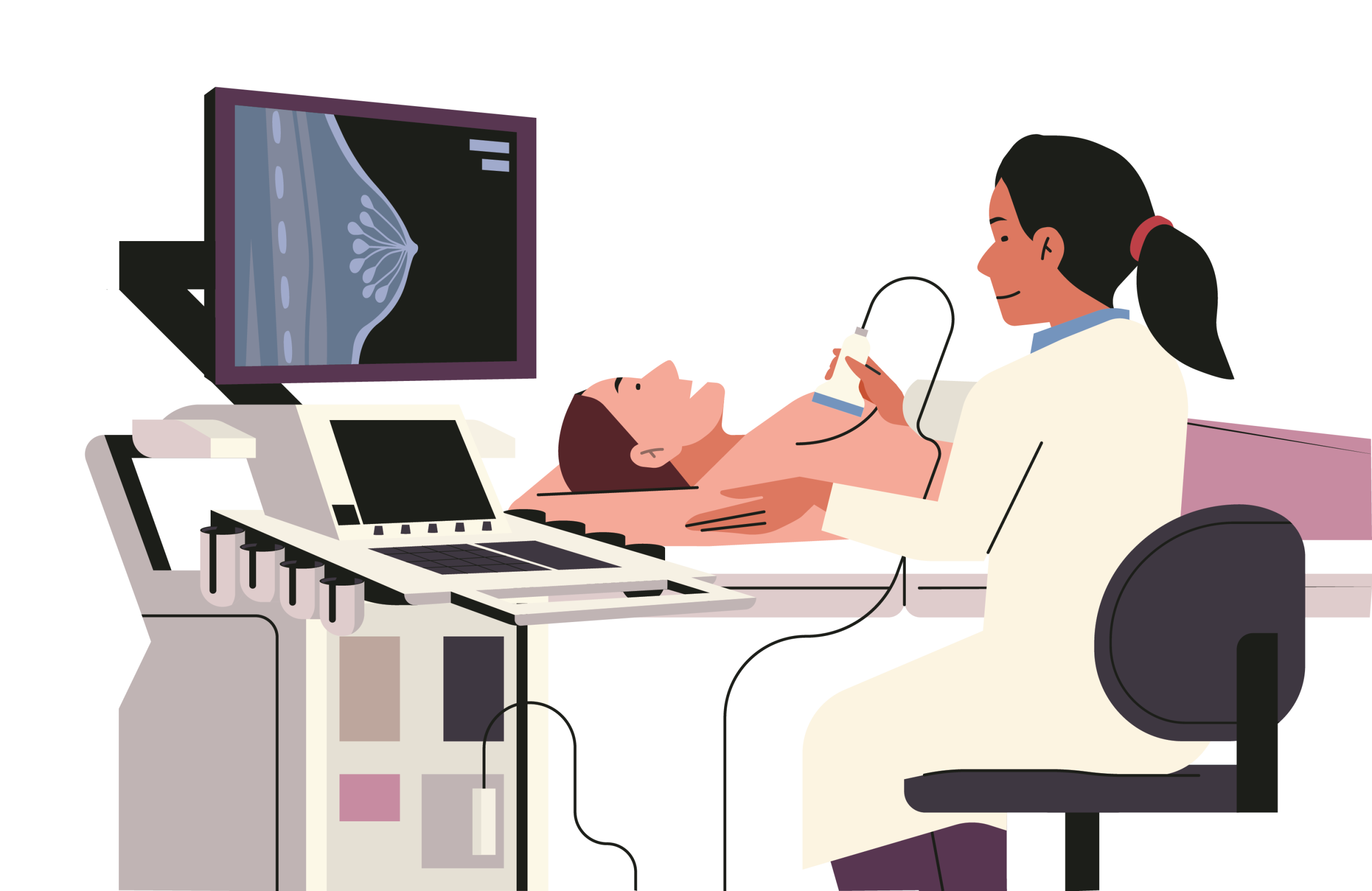
Ultrasound is overall the preferred imaging modality for breast biopsies, mainly because it is more comfortable for the patient. Most biopsies will be done under ultrasound if they can, but if the lesion is not visible under ultrasound (like small microcalcification clusters) then it will need to be performed with mammography.
After the tissue has been taken, the clinician will likely put in something called a marker clip. This tiny (usually metal) marker will be placed exactly where they took the biopsy from, so that in future if they want to keep track of the area it can easily be identified in scans.
The metal clip in the markers in our portfolio at It’s Interventional are 1-4mm in size – seriously tiny! You shouldn’t be able to feel it, you can still have an MRI and it won’t set off the metal detector at the airport.
Other Questions?
For 24 hours after a biopsy, you should: Take it easy! Avoid strenuous exercise, leave your dressing on, try not to get it soaking wet (a shower is fine, but don’t swim or soak in the bath), and avoid any heavy lifting. If you experience any pain, take over-the-counter painkillers.
If you have any excessive swelling or redness, the breast becomes hot to the touch or bleeds a lot, you should consult the breast unit or your GP over the phone. Outside of working hours, it may require checking in A&E.
While mammographers will always be female, there are male radiologists that do perform some ultrasound scans, biopsies and physical examinations. You will always have a female chaperone however, so you will never be alone with a man. If you are not comfortable with seeing a male clinician, you may of course request to only be seen by female staff. Do bear in mind though that this could mean having to come back another day to complete your testing or possibly even require you to travel to a different site. To do this, Becky recommends calling your breast unit prior to your appointment and they will do their best to accommodate your needs


